Materials around the peptide
KPV in hydrogels and nanoparticles
Once incorporation becomes a physical question, the material surrounding a peptide deserves attention too.
KPV held in a gel
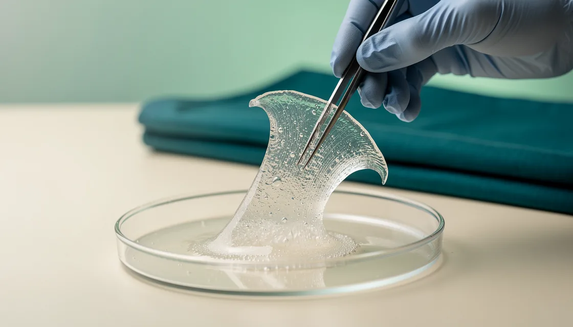
A materials-characterization study used a self-cross-linked hydrogel to stabilize KPV and enable controlled release. A hydrogel is a water-containing network; here, building that network around the peptide was part of the experimental work. The finding concerns a material and its handling of KPV.
A separate double-network hydrogel study in mice reported restoration of the gut mucosal barrier and reduced inflammation in an inflamed-colon model. That goes further into biology: a KPV-binding material was studied in a living animal with colitis.
I’d been treating the surrounding material as a minor detail. Stabilization and release deserve a much larger share of my attention after these experiments. Stabilizing a peptide, releasing it and observing a tissue response are connected tasks, and the formulation can change what is actually being tested.
A colon-targeted formulation
In a mouse DSS-colitis study, KPV-loaded nanoparticles were functionalized with hyaluronic acid and embedded in a chitosan/alginate hydrogel for oral delivery. The targeted formulation showed stronger prevention of mucosal damage and downregulation of TNF-alpha than non-targeted delivery, with faster mucosal healing.
The hyaluronic acid, nanoparticle and gel belong in any description of that result. Removing them from the story would silently replace the tested preparation with a much simpler one. The experiments make the labour around KPV remarkably visible.
This comparison also answers a different question from the intestinal uptake experiment. The uptake work identifies a cellular transport route. The formulation work tests an assembled delivery system in mice. Both concern arrival, at different scales.
KPV combined with FK506
A PepT1-targeted preparation combining KPV with the immunosuppressant FK506 improved outcomes in acute and chronic DSS-colitis models in mice. The study reported restoration of tight-junction proteins and lower inflammatory cytokines beyond either agent alone. Tight junctions are the protein structures that help adjacent epithelial cells seal together.
I admire the inclusion of both individual agents in this comparison. Still, the resulting preparation contains both agents in a co-assembled nanodrug. Its effect belongs to that tested combination and mouse setting.
The larger temptation is to read all these materials papers as a single accumulating endorsement of KPV. A collective verdict would erase the experimental differences: several preparations have produced specific results in different experiments. Their ingenuity is worth discussing without turning the formulations into instructions for personal use.